44 microscope labeled diagram
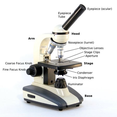
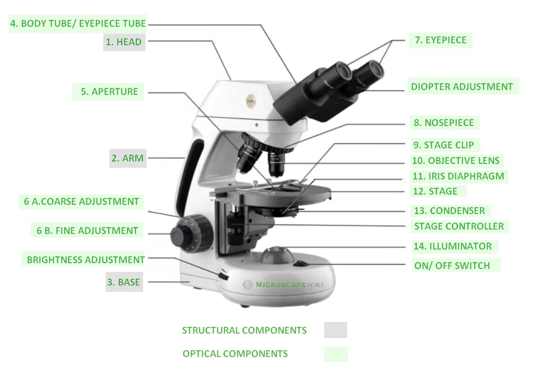
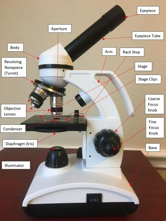
17 Parts of a Microscope with Functions and Diagram Jul 29, 2022 ... References. microscope | Types, Parts, History, Diagram, & Facts | BritannicaParts of the Microscope with Labeling (also Free Printouts) – ... Compound Microscope Parts - Labeled Diagram and their Functions Labeled diagram of a compound microscope Major structural parts of a compound microscope There are three major structural parts of a compound microscope. The head includes the upper part of the microscope, which houses the most critical optical components, and the eyepiece tube of the microscope.
Light Microscope- Definition, Principle, Types, Parts, Labeled Diagram ... Figure: Labeled Diagram of a Light Microscope. Types of light microscopes (optical microscope) With the evolved field of Microbiology, the microscopes. used to view specimens are both simple and compound light microscopes, all using lenses. The difference is simple light microscopes use a single lens for magnification while compound lenses use ...
Microscope labeled diagram
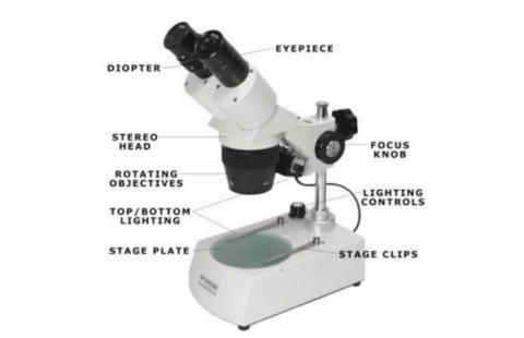
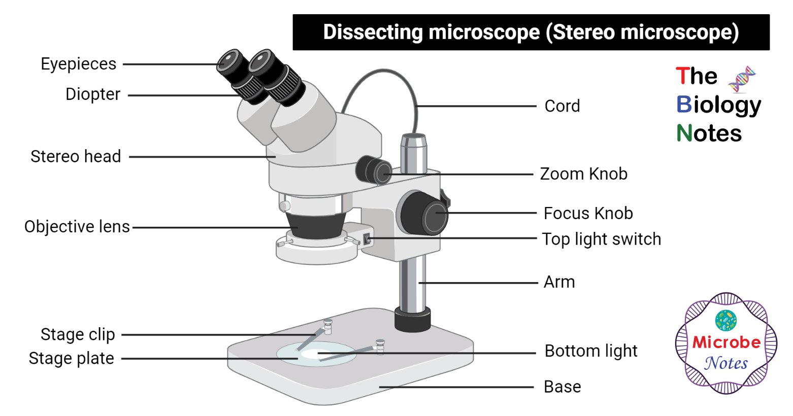
Parts of Stereo Microscope (Dissecting microscope) - labeled diagram ... Labeled part diagram of a stereo microscope Major structural parts of a stereo microscope. There are three major structural parts of a stereo microscope. The viewing Head includes the upper part of the microscope, which houses the most critical optical components, including the eyepiece, objective lens, and light source of the microscope. Compound Microscope Parts, Functions, and Labeled Diagram Compound Microscope Parts, Functions, and Labeled Diagram - New York Microscope Company Microscope Experts Since 1979 (877) 877-7274 Request a Quote Contact Us Sign in USD Cart ( 0 ) Compound Microscopes Stereo Microscopes Digital Microscopy Applications Accessories Brands Services Shop PPE Microscope Types (with labeled diagrams) and Functions Electron microscope labeled diagram The different types of electron microscopes are: Transmission Electron Microscope Scanning Electron Microscope Reflection Electron Microscope Scanning transmission electron microscope Scanning tunneling microscopy Electron microscope functions: Semiconductors and Data Storage Industry Failure Analysis
Microscope labeled diagram. Microscope Parts, Types & Diagram | What is a Microscope? Microscope Diagram There are many illustrations available for the diagram of a light microscope. The essential parts include the head, base, arms, lenses, and lights. In diagrams, one... Microscopy: Intro to microscopes & how they work (article) - Khan Academy Magnification is a measure of how much larger a microscope (or set of lenses within a microscope) causes an object to appear. For instance, the light microscopes typically used in high schools and colleges magnify up to about 400 times actual size. So, something that was 1 mm wide in real life would be 400 mm wide in the microscope image. Acoustic Electric Guitar Wiring Diagram Labeled Microscope Fiso El Musica. June 13, 2022 Uncategorized Comments Off. Sims raises his music game with the release of a new Amapiano banger dubbed "Balele" featuring Fiso El Musica. The star singer has released a couple of songs recently, "Balele" stands out from that lot. [nextmp3-generate keyword="Sims - Balele feat. label microscope diagram | Charts - Pinterest Light microscope, optical microscope diagrams. Label microscope diagram. Microscope labeled diagram. Microscope lens. Less.
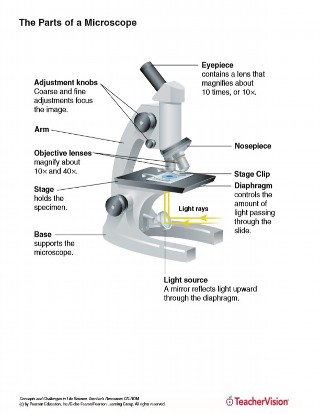
Microscope Parts and Functions Microscope Parts and Functions With Labeled Diagram and Functions How does a Compound Microscope Work?. Before exploring microscope parts and functions, you should probably understand that the compound light microscope is more complicated than just a microscope with more than one lens.. First, the purpose of a microscope is to magnify a small object or to magnify the fine details of a larger ... 16 Essential Microscope Parts: Names, Functions & Labeled Diagram Condenser. The condenser is to focus the light, which passes from the microscopic illuminator to the specimen. This condenser is located just below the diaphragm. This diaphragm is one of the essential parts of the compound microscope, which will help to get an accurate and sharp image. The condenser has a magnification power of 400X and above. Parts of a microscope with functions and labeled diagram Figure: Diagram of parts of a microscope There are three structural parts of the microscope i.e. head, base, and arm. Head - This is also known as the body. It carries the optical parts in the upper part of the microscope. Base - It acts as microscopes support. It also carries microscopic illuminators. Stratified Squamous Epithelium Under Microscope with Labeled Diagram ... The labeled diagrams help you identify the keratinized and nonkeratinized stratified squamous epithelium under the light microscope. You will see the significant difference in the cell layers and the presence or absence of keratin in the superficial layer between keratinized and nonkeratinized stratified squamous epithelium.
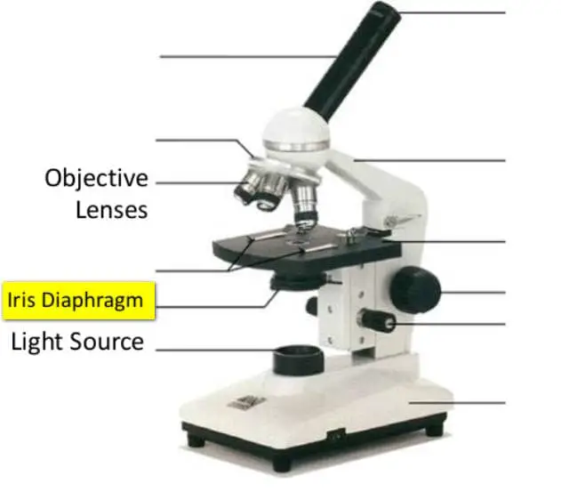
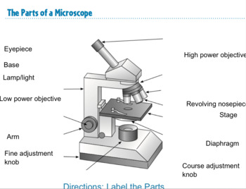
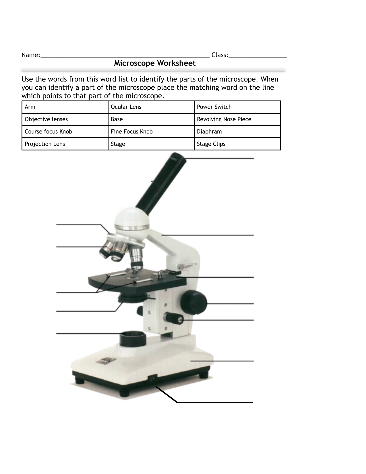
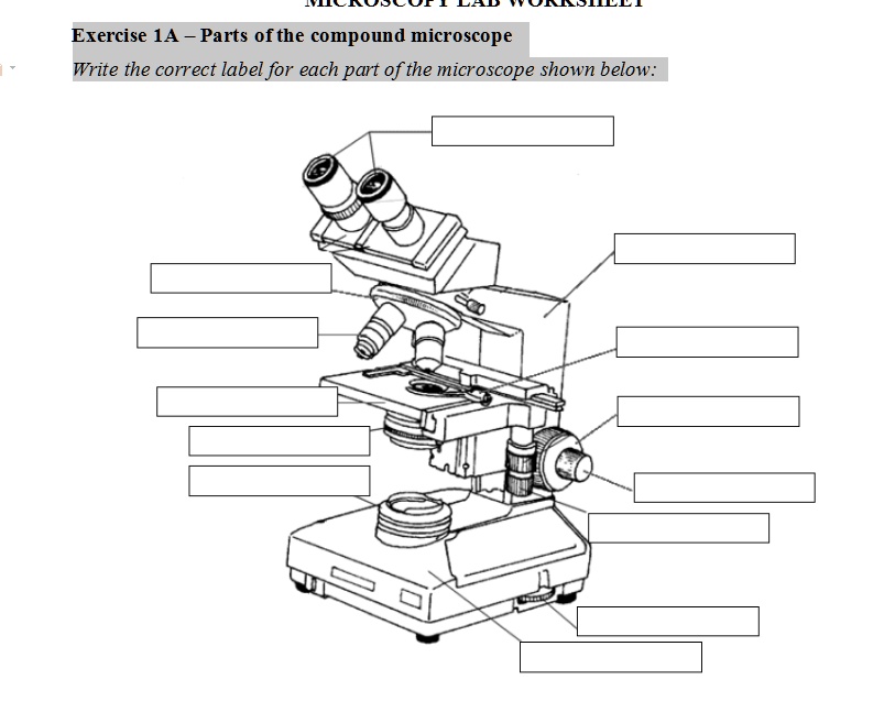
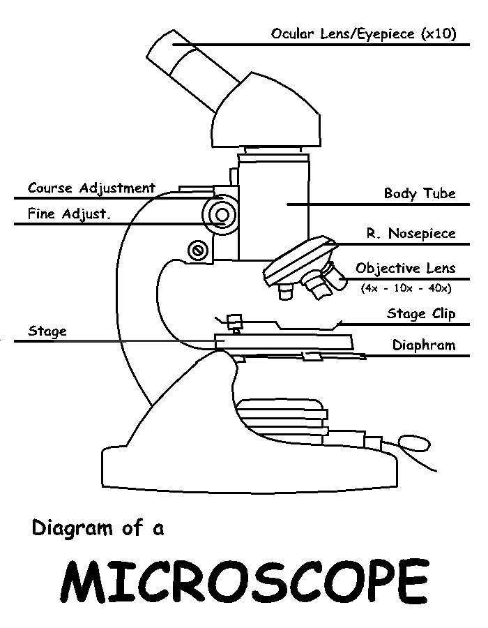
A Study of the Microscope and its Functions With a Labeled Diagram ... These labeled microscope diagrams and the functions of its various parts, attempt to simplify the microscope for you. However, as the saying goes, 'practice makes perfect', here is a blank compound microscope diagram and blank electron microscope diagram to label. Download the diagrams and practice labeling the different parts of these ... Labeling the Parts of the Microscope Labeling the Parts of the Microscope. This activity has been designed for use in homes and schools. Each microscope layout (both blank and the version with ... Cardiac Muscle Under Microscope with Labeled Diagram Here, I tried to show you all the important microscope features of the cardiac myocytes and fibers with the 40X, 100X, and 400X labeled diagrams. Cardiac Muscle Microscope Slide 40X First, let's see the diagram of the cardiac muscle microscope slide with 40X. Parts of the Microscope (Labeled Diagrams) Simple microscope labelled diagram Image created with Biorender Tube/Body Tube It serves as the connector between the eyepiece/ocular and objective lenses. Objective lenses The lenses have varying magnifying power, which typically consists of 10x, 40x, and 100x.
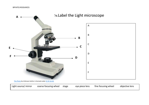
Label the microscope - Science Learning Hub In this interactive, you can label the different parts of a microscope. Use this with the Microscope parts activity to help students identify and label the main parts of a microscope and then describe their functions. Drag and drop the text labels onto the microscope diagram.
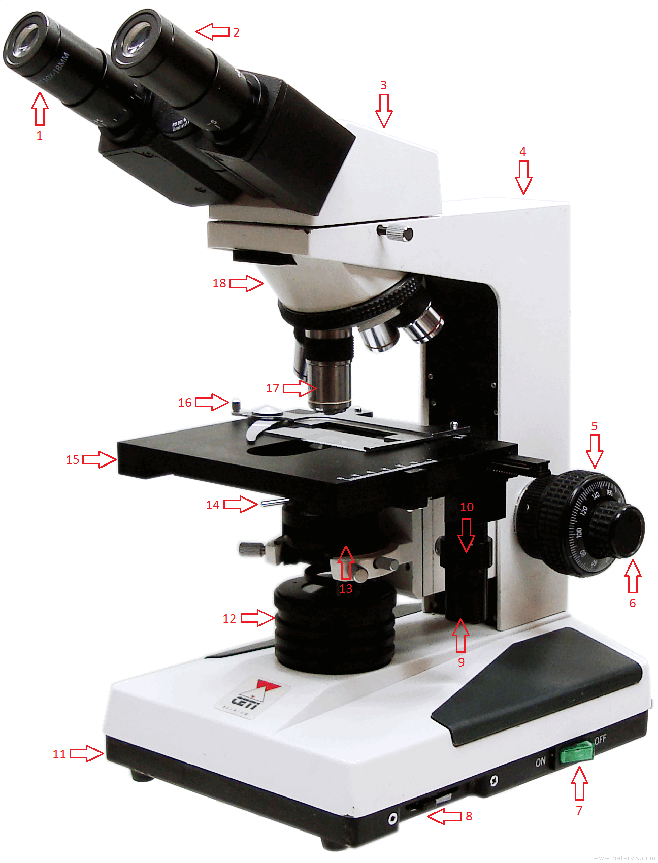
Binocular Microscope Anatomy - Parts and Functions with a Labeled Diagram Let's see the microscope labeled diagram; you will find the flat platform where the slide is placed. Again, this microscope stage lies perpendicular to the optical system or pathway. In some microscopes, the stage can move when the focus is adjusted. Again, the microscope stage is often designed with mechanical devices for holding and moving ...
Types of Microscopes: Definition, Working Principle, Diagram ... There are also microscope types that find application in metallurgy and studying three-dimensional samples. In this article, there are 5 such microscope types that are discussed along with their diagram, working principle and applications. These five types of microscopes are: Simple microscope. Compound microscope.
Skeletal Muscle Under Microscope with Labeled Diagram The provided labeled diagrams identify all these microscopic features from the skeletal muscle histology slide. You might see these microscopic features of skeletal muscle both from the longitudinal and cross (transverse) sections. Summary of the skeletal muscle microscope slide
Parts of a microscope with functions and labeled diagram - The Compound ... Parts off a microscope with functions and labeling plot. Jay 13, 2023 September 17, 2022 by Confidence Mokobi September 17, 2022 by Confidence Mokobi
Microscope labeled diagram - SlideShare Microscope labeled diagram 1. The Microscope Image courtesy of: Microscopehelp.com Basic rules to using the microscope 1. You should always carry a microscope with two hands, one on the arm and the other under the base. 2. You should always start on the lowest power objective lens and should always leave the microscope on the low power lens ...
Simple Columnar Epithelium Under a Microscope with Labeled Diagram ... Now, the labeled diagram of simple columnar epithelium under a microscope shows two types of cells - Columnar cells with microvilli or brush border, and Oval-shaped goblet cells, Here, the apical microvilli or brush border from the surface look like the reddish outer layer with longitudinal striation.
Compound Microscope Parts – Labeled Diagram and their Functions Jan 27, 2021 - Microscope parts include eyepiece (10x), objective lenses (4x, 10x, 40x, 100x), fine and coarse focus, slide holder, condenser, ...
Microscope Parts & Specifications Labeled Diagram Learn about a microscopes parts and its functions including the eyepiece, objectives, and condenser with our labeled diagram.
Microscope Types (with labeled diagrams) and Functions Electron microscope labeled diagram The different types of electron microscopes are: Transmission Electron Microscope Scanning Electron Microscope Reflection Electron Microscope Scanning transmission electron microscope Scanning tunneling microscopy Electron microscope functions: Semiconductors and Data Storage Industry Failure Analysis
Compound Microscope Parts, Functions, and Labeled Diagram Compound Microscope Parts, Functions, and Labeled Diagram - New York Microscope Company Microscope Experts Since 1979 (877) 877-7274 Request a Quote Contact Us Sign in USD Cart ( 0 ) Compound Microscopes Stereo Microscopes Digital Microscopy Applications Accessories Brands Services Shop PPE
Parts of Stereo Microscope (Dissecting microscope) - labeled diagram ... Labeled part diagram of a stereo microscope Major structural parts of a stereo microscope. There are three major structural parts of a stereo microscope. The viewing Head includes the upper part of the microscope, which houses the most critical optical components, including the eyepiece, objective lens, and light source of the microscope.














Komentar
Posting Komentar